Exploring Blood Cells Under a Microscope: A Visual Guide

<!DOCTYPE html>
Have you ever wondered what blood cells look like up close? Exploring blood cells under a microscope opens a fascinating window into the microscopic world of our circulatory system. Whether you’re a student, a medical professional, or simply curious, this guide will walk you through the process of observing and understanding the different types of blood cells. From red blood cells to white blood cells and platelets, each plays a crucial role in maintaining health. Let’s dive into the steps, tools, and techniques needed to explore blood cells under a microscope, (blood cell microscopy, hematology, laboratory techniques)
Essential Tools for Blood Cell Microscopy

Before you begin, ensure you have the right tools for a successful observation. Here’s what you’ll need:
- Microscope: A compound microscope with at least 400x magnification.
- Slides and Cover Slips: Clean, high-quality glass slides and cover slips.
- Blood Sample: A small drop of blood, preferably from a finger prick.
- Stains (Optional): Wright’s or Giemsa stain for better visualization of cell structures.
- Pipettes and Spreaders: For precise blood sample handling.
📌 Note: Always handle blood samples with care and follow safety protocols to avoid contamination.
Step-by-Step Guide to Preparing a Blood Smear

Step 1: Collect the Blood Sample
Start by cleaning the fingertip with alcohol. Use a sterile lancet to prick the finger and collect a small drop of blood. (blood sample collection, sterile techniques)
Step 2: Prepare the Slide
Place a clean glass slide on a flat surface. Using a pipette, transfer a small drop of blood to one end of the slide. (microscope slide preparation, pipetting techniques)
Step 3: Create the Smear
Hold a spreader at a 30-degree angle and gently push the blood drop across the slide to create a thin, even smear. Allow it to air dry completely. (blood smear technique, slide preparation)
Step 4: Stain the Slide (Optional)
If using a stain, carefully apply it to the dried smear and follow the manufacturer’s instructions for fixation and washing. (blood cell staining, hematology stains)
Observing Blood Cells Under the Microscope

Once your slide is ready, it’s time to examine it under the microscope. Here’s how:
- Focus on Low Power: Place the slide on the microscope stage and start with the lowest magnification to locate the smear.
- Switch to High Power: Gradually increase the magnification to 400x or higher for detailed observation.
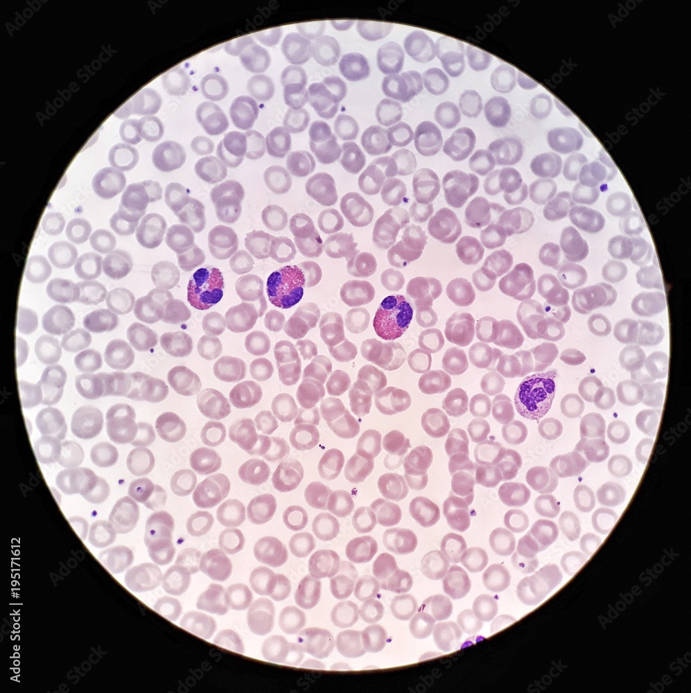
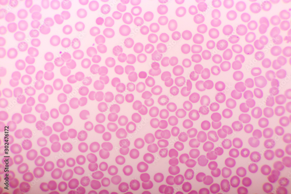
- Identify Cell Types: Look for distinctive features of red blood cells (disc-shaped), white blood cells (larger, with nuclei), and platelets (small, irregular shapes).
| Cell Type | Shape | Size | Function |
|---|---|---|---|
| Red Blood Cells | Biconcave disc | 6-8 μm | Carry oxygen |
| White Blood Cells | Irregular | 10-15 μm | Fight infections |
| Platelets | Irregular | 2-3 μm | Aid in clotting |

Tips for Better Microscopic Observation

- Lighting: Ensure proper illumination for clear visibility.
- Cleanliness: Keep slides and lenses free from dust and fingerprints.
- Practice: Regular practice improves your ability to identify cell types accurately.
Exploring blood cells under a microscope is not only educational but also a rewarding experience. Whether for academic purposes or personal curiosity, understanding the basics of blood cell microscopy can deepen your appreciation for the complexity of human biology. (microscopic observation, blood cell identification, educational microscopy)
What magnification is best for observing blood cells?
+A magnification of 400x or higher is ideal for detailed observation of blood cells.
Can I observe blood cells without staining?
+Yes, but staining enhances visibility and makes it easier to distinguish cell types.
How do I safely handle blood samples?
+Use sterile tools, wear gloves, and dispose of samples properly to avoid contamination.


