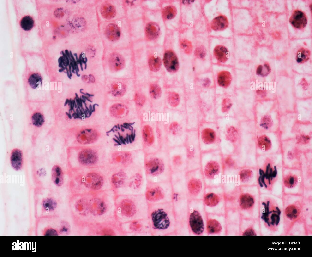
Unveiling Mitosis in Allium Root Tip: A Microscopic Journey

Exploring the intricate process of mitosis in Allium root tip offers a fascinating glimpse into the fundamental mechanisms of cell division. Through a microscopic journey, we can observe how cells replicate, ensuring growth and repair in organisms. This blog delves into the steps of preparing and examining Allium root tip cells, highlighting their significance in biology and research. Whether you're a student, educator, or enthusiast, understanding mitosis in this context enhances your grasp of cellular biology, (cell division, microscopic analysis, Allium root tip preparation).
Why Study Mitosis in Allium Root Tip?

Allium root tips, such as those from onions or garlic, are ideal for studying mitosis due to their rapid cell division. The root tip meristem contains cells in various stages of mitosis, making it a perfect model for observation. This study is crucial for understanding how organisms grow and develop, (root tip meristem, cell division stages, biological research).
Materials Needed for Observing Mitosis

Before embarking on this microscopic journey, gather the following materials:
- Allium root tip (onion or garlic)
- Microscope with high magnification
- Stain (e.g., acetic orcein or Feulgen stain)
- Blade and slides
- Hydrochloric acid and alcohol for fixation
📌 Note: Ensure all tools are clean to avoid contamination during preparation, (microscope preparation, staining techniques, laboratory tools).
Step-by-Step Guide to Preparing Allium Root Tip

Step 1: Collect and Prepare the Root Tip
Carefully remove the Allium root tip, ensuring it remains intact. Trim it to 5–8 mm for optimal observation, (root tip collection, sample preparation, microscopy techniques).
Step 2: Fixation Process
Fix the root tip in a solution of hydrochloric acid and alcohol (1:3 ratio) for 5–10 minutes. This stops cell division and preserves the sample, (fixation process, cell preservation, laboratory procedures).
Step 3: Hydrolysis and Staining
Hydrolyze the sample in 45% acetic acid for 5 minutes, then stain it with acetic orcein for 10–15 minutes. Staining enhances visibility under the microscope, (hydrolysis, staining process, cellular visibility).
Step 4: Slide Preparation and Observation
Place the stained root tip on a slide, add a coverslip, and gently press to spread the cells. Observe under a microscope, focusing on the metaphase stage for clear chromosome alignment, (slide preparation, microscopic observation, metaphase stage).
| Stage | Key Features |
|---|---|
| Prophase | Chromosomes condense, nuclear envelope breaks down |
| Metaphase | Chromosomes align at the equatorial plate |
| Anaphase | Chromosomes separate and move to opposite poles |
| Telophase | Nuclear envelope reforms, chromosomes decondense |

Observing mitosis in Allium root tip provides invaluable insights into cellular replication and growth. By following these steps, you can successfully prepare and examine root tip cells, enhancing your understanding of biological processes. This microscopic journey not only educates but also inspires further exploration into the wonders of cell biology, (cellular replication, biological processes, educational microscopy).
Why is Allium root tip ideal for studying mitosis?
+
Allium root tips have a high rate of cell division, making it easy to observe all stages of mitosis under a microscope, (cell division, microscopic analysis, Allium root tip preparation).
What is the purpose of staining in this experiment?
+
Staining enhances the visibility of chromosomes and cell structures, making it easier to identify different stages of mitosis, (staining techniques, cellular visibility, laboratory tools).
Can other plants be used for this experiment?
+
Yes, other plants with actively growing root tips, such as beans or wheat, can also be used, though Allium is preferred for its clarity and ease of preparation, (plant alternatives, root tip collection, microscopy techniques).


