Exploring the Human Body Through Pictures

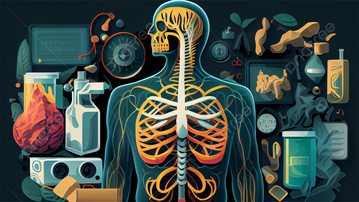
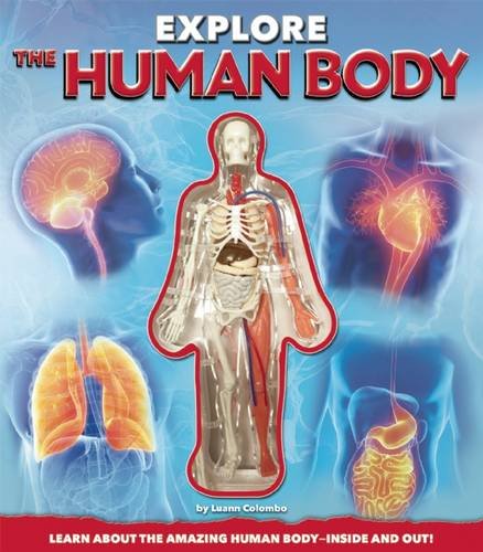
The human body is a marvel of complexity, with intricate systems working in harmony to sustain life. From the microscopic cells to the visible organs, every part plays a crucial role. Exploring the human body through pictures offers a unique and accessible way to understand its anatomy, functions, and the fascinating interplay of its components. Whether you’re a student, a medical professional, or simply curious, visual aids can make learning both engaging and informative. (human anatomy, medical illustrations, visual learning)
Why Use Pictures to Explore the Human Body?

Visual learning is a powerful tool that enhances comprehension and retention. Pictures of the human body provide a clear, detailed view of anatomical structures, making it easier to grasp complex concepts. Medical illustrations, diagrams, and 3D models bridge the gap between theory and practice, allowing learners to visualize how different systems function together. (anatomical diagrams, 3D models, visual comprehension)
Key Areas to Explore Through Visuals

The Skeletal System
The skeletal system is the body’s framework, providing support and protection. Pictures of the skeletal system highlight the arrangement of bones, joints, and cartilage. Visuals can help identify the function of each bone, such as the femur (thigh bone) as the strongest bone in the body. (skeletal system, bone structure, femur)
The Muscular System
Muscles are essential for movement, posture, and even internal functions like digestion. Images of the muscular system showcase the layers of muscles, their attachments to bones, and their roles in various actions. For instance, the biceps and triceps work together to allow arm movement. (muscular system, biceps, triceps)
The Circulatory System
The circulatory system is a network of blood vessels and the heart, responsible for transporting oxygen and nutrients. Visual representations of this system illustrate how blood flows through arteries, veins, and capillaries, ensuring every cell receives essential resources. (circulatory system, heart, blood vessels)
Tools and Resources for Visual Exploration

Anatomy Atlases
Anatomy atlases are comprehensive collections of high-quality images, diagrams, and labels. They are invaluable for students and professionals alike, offering detailed insights into the body’s structure. (anatomy atlas, medical textbooks, detailed diagrams)
Interactive 3D Models
Interactive 3D models allow users to rotate, zoom, and dissect virtual representations of the human body. These tools are particularly useful for understanding spatial relationships between organs and systems. (3D anatomy models, virtual dissection, spatial relationships)
Medical Imaging Techniques
Techniques like X-rays, MRIs, and CT scans provide real-life visuals of the body’s internal structures. These images are crucial for diagnosis and treatment planning in medical practice. (medical imaging, X-rays, MRI scans)
📌 Note: When using medical imaging, ensure proper training to interpret results accurately.
Benefits of Visual Learning in Anatomy

- Enhanced Understanding: Visuals simplify complex anatomical concepts.
- Better Retention: Images improve memory and recall of information.
- Engagement: Interactive tools make learning more enjoyable and effective.
Checklist for Exploring the Human Body Through Pictures

- Choose high-quality, accurate visual resources.
- Use labeled diagrams for clarity.
- Explore interactive tools for hands-on learning.
- Combine visuals with textual explanations for comprehensive understanding.
Exploring the human body through pictures is an effective and engaging way to learn about its intricate design. From anatomy atlases to interactive 3D models, visual tools cater to diverse learning styles and needs. By leveraging these resources, anyone can gain a deeper appreciation for the body’s complexity and functionality. Whether for education, medical practice, or personal curiosity, visual exploration opens up a world of knowledge. (visual exploration, anatomy education, medical practice)
What are the best resources for visual anatomy learning?
+Anatomy atlases, interactive 3D models, and medical imaging techniques like MRIs and CT scans are excellent resources for visual anatomy learning.
How do pictures improve understanding of the human body?
+Pictures simplify complex anatomical structures, enhance memory retention, and make learning more engaging and accessible.
Can visual tools replace traditional anatomy textbooks?
+While visual tools are highly effective, they complement traditional textbooks by providing a more interactive and dynamic learning experience.


