Spinal Cord Cross Section: Labeled Anatomy Guide
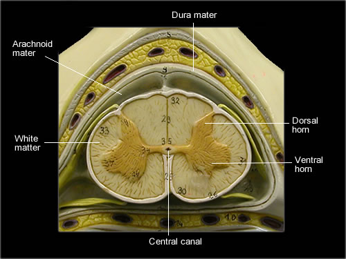
Understanding the spinal cord’s anatomy is crucial for medical professionals, students, and anyone interested in the human body’s intricate structure. A spinal cord cross section reveals the complex organization of this vital structure, which plays a key role in transmitting signals between the brain and the rest of the body. In this guide, we’ll explore the labeled anatomy of a spinal cord cross section, highlighting its key components and functions. (spinal cord anatomy, spinal cord cross section, labeled anatomy guide)
Key Components of a Spinal Cord Cross Section
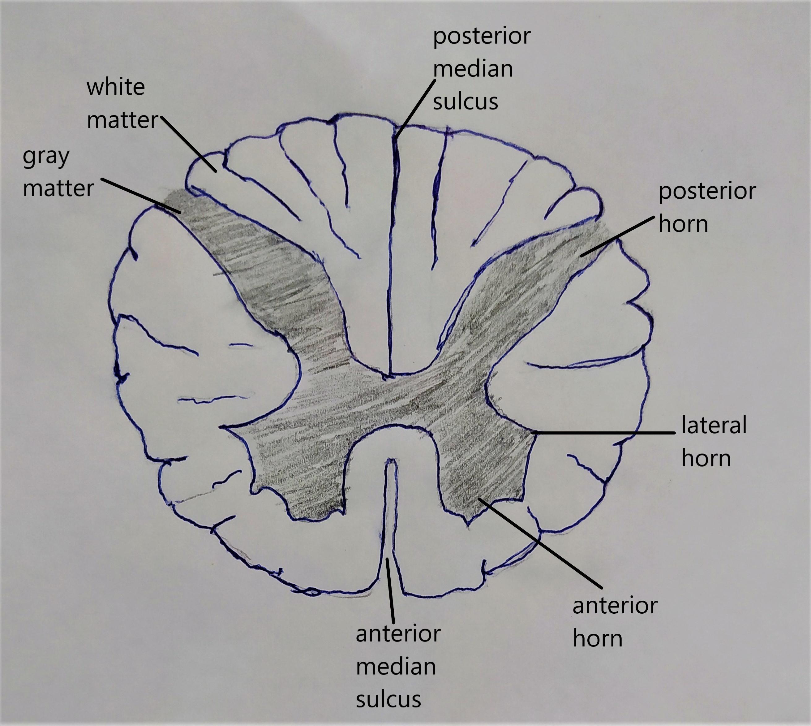
When examining a spinal cord cross section, several distinct regions become apparent. These include the gray matter and white matter, each serving unique functions. The gray matter is butterfly-shaped and consists of cell bodies, synapses, and unmyelinated axons. It’s responsible for processing information and relaying signals. The white matter, on the other hand, is composed of myelinated axons that transmit signals quickly over long distances. (gray matter, white matter, spinal cord function)
Gray Matter: The Processing Center
The gray matter is divided into several regions, including the anterior horns, posterior horns, and lateral horns. Each region contains specific types of neurons that contribute to different functions, such as motor control and sensory processing. (anterior horns, posterior horns, lateral horns)
| Region | Function |
|---|---|
| Anterior Horns | Motor neuron cell bodies |
| Posterior Horns | Sensory neuron cell bodies |
| Lateral Horns | Sympathetic neuron cell bodies (thoracic and lumbar regions) |
📌 Note: The lateral horns are only present in the thoracic and lumbar regions of the spinal cord.
White Matter: The Communication Highway
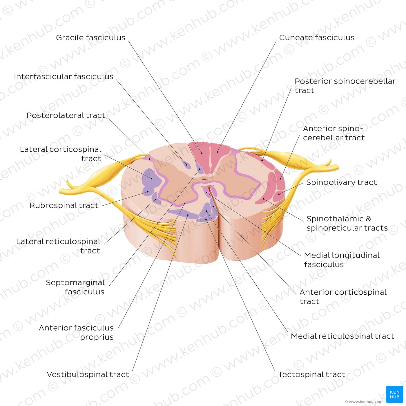
The white matter is organized into ascending tracts and descending tracts. Ascending tracts transmit sensory information from the body to the brain, while descending tracts carry motor commands from the brain to the body. (ascending tracts, descending tracts, spinal cord communication)
Spinal Cord Cross Section: Labeled Anatomy

A labeled spinal cord cross section typically includes the following structures:
- Central Canal: Filled with cerebrospinal fluid, providing cushioning and nutrients.
- Anterior Median Fissure: A groove on the anterior surface of the spinal cord.
- Posterior Median Sulcus: A groove on the posterior surface of the spinal cord.
- Spinal Nerve Roots: Exit the spinal cord through the intervertebral foramina.
(central canal, anterior median fissure, posterior median sulcus, spinal nerve roots)
Practical Applications and Commercial Solutions
For those seeking spinal cord models or anatomical charts, numerous options are available. High-quality models can aid in medical education, patient consultation, and research. (spinal cord models, anatomical charts, medical education)
To summarize, understanding the spinal cord cross section is essential for grasping the complexities of human anatomy. By familiarizing yourself with its key components and functions, you’ll be better equipped to appreciate the spinal cord’s role in maintaining overall health and function. (spinal cord health, human anatomy, medical knowledge)
What is the primary function of the spinal cord?
+The spinal cord’s primary function is to transmit signals between the brain and the rest of the body, enabling movement, sensation, and autonomic functions.
How is the gray matter organized in the spinal cord?
+The gray matter is butterfly-shaped and divided into regions such as the anterior horns, posterior horns, and lateral horns, each serving specific functions.
What are ascending and descending tracts in the spinal cord?
+Ascending tracts transmit sensory information from the body to the brain, while descending tracts carry motor commands from the brain to the body.

